According to the American Veterinary Dental Society, approximately 80% of dogs and 70% of cats over 3 years of age have some degree of periodontal disease. Despite this alarming statistic, most pet owners are unaware of the importance of oral care in their pets, nor do they realize that it can be linked to other health related issues.
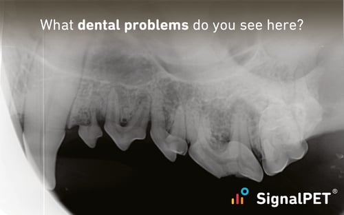
All patients benefit from the information provided by dental radiographs. As the statistics confirm, a majority of patients will have some form of oral disease at some point in their lives. Pet oral care is important to the overall health of the patient and regular dental radiographs are an essential part of that care. Radiographs help track healthy dental maintenance, detect potential problems, and/or help guide a proper treatment plan.
At SignalPET, we recognize the importance of dental care in your practice. We are excited to announce the upcoming release of SignalSMILE and its range of dental tests. Your second set of eyes on dental films has arrived.
How SignalSMILE Can Help
SignalPET is continually training its artificial intelligence (AI) to identify the most common dental pathologies seen in your radiographs as part of your routine dental procedures or COHATs (comprehensive oral health assessment and treatment). With SignalSMILE, you will see these pathologies show up under five separate tests, each of which includes an outline functionality to highlight the pathology in the image.
Severe Attachment/Alveolar Bone Loss
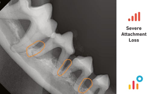
Periodontal disease has been identified as the most common disease found in adult dogs and cats. The main indicators of periodontal disease are inflammation and alveolar bone loss. If present in the patient, periodontal disease requires medical attention and monitoring. Left untreated, periodontal disease will progress and lead to eventual loss of affected teeth. Alveolar bone loss can occur in vertical and horizontal planes, both of which are recognized in the Severe Attachment Loss test. This particular test assists in differentiating the four grades of periodontal disease by indicating the severity of attachment loss with red or green bars. Grade 4 periodontal disease is characterized by greater than 50% attachment loss and is reflected in a test result of four red bars. Two red bars coordinate with grade 3 periodontal disease or 25-50% attachment loss, two green bars with grade 2 periodontal disease or 1-25% attachment loss, four green bars with no apparent attachment loss.
Tooth Fracture
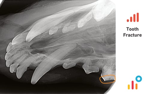
Fractured teeth are a common occurrence in veterinary practice and are often caused by external trauma or by chewing hard objects (bones, rocks, antlers, some chew toys, etc.). Fractures can also be secondary to pathology within the tooth, or can even be caused by a veterinarian’s own attempts at extraction (i.e. retained roots). Regardless of the causation or type of fracture present, fractured teeth require attention. The Tooth Fracture test is able to identify fractures that may be uncomplicated or complicated, and is able to identify them regardless of location - root or crown. When deciding on a treatment plan for a fractured tooth, multiple factors must be taken into account, with radiographic interpretation being one of the key factors.
Furcation Bone Loss
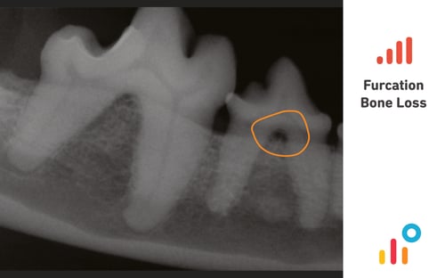
Periodontal disease that extends to the furcation of a tooth can lead to the loss of alveolar bone in this region. The furcation is a normal anatomical region where the roots begin to diverge in a multirooted tooth and is normally filled with bone. If furcation disease is present, the stage of disease should be determined by performing an oral exam under anesthesia along with dental radiographs. When bone loss at the furcation is apparent on radiographs, it is an indication that severe disease is present. The Furcation Bone Loss test identifies radiolucency at the furcation of a tooth that is indicative of loss of alveolar bone in this region. Teeth identified with this test will require medical attention.
Tooth Resorption
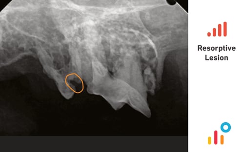
Tooth resorption is a progressive destructive process by which the dentin of affected teeth erodes and eventually becomes irreparably destroyed by clastic cells. Despite the high prevalence in cats (20-72%) and dogs (17.9-53.6%), much confusion exists with regards to its nomenclature, etiology, classification, diagnosis and treatment plan. Radiology is essential in diagnosing tooth resorption. According to one report, intraoral radiographs revealed pathology in 42% of cats and 28% of dogs when no pathology was noted on the oral examination; in patients with abnormal findings, radiology revealed additional pathology in 50% of dogs and 54% of cats. Depending on the type of resorption present, resorption can appear as radiolucency in the crown and/or root, focal loss of the periodontal ligament detail, focal enlargement of the root canal, and/ or varying degrees of disappearance of the root. The Resorptive Lesion test searches for radiological findings associated with tooth resorption and outlines the area of concern. Tooth resorption can be an extremely painful process. As such, current treatment plans are directed to relieving discomfort and pain, and often limited to tooth extraction or crown amputation.
Periapical Lucencies
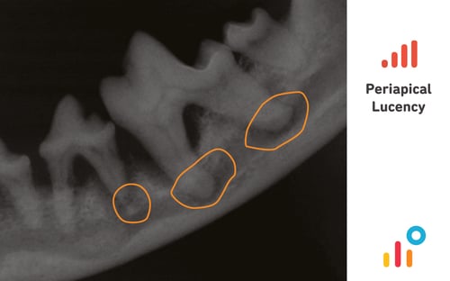
Periapical lucencies are dark regions located at the root apex and may be pathological or non-pathological. Non pathologic lucencies are often referred to as chevron lesions whereas pathological lucenences represent lesions of endodontic origin (LEO). The common differentials include granuloma, cyst, or abscess and/or neoplasia. Pathological periapical lucencies tend to be circular, centred over the apex of the root, large and/or irregular shaped. The Periapical Lucency test searches for radiological characteristics of pathological lucencies and demarcates the area of concern.
Significant Tooth with Pathology
An additional alert provided by SignalSMILE is the Significant Tooth with Pathology notification. This alerts you to the presence of pathology (identified by one or more of the five tests) in a carnassial or canine tooth. These specific teeth deserve special attention and treatment considerations. Responsible for tearing, grinding, chewing or crushing, the canines and carnassials serve a more significant function within the mouth of our companion animals and thus have more anchoring into alveolar bone. This alert serves as a reminder that treatment of these teeth should be approached with more caution than other teeth.