Hacking Feline Asthma - It ain't easy being wheezy
Published by SignalPET on January 22, 2021

Feline chronic bronchitis (commonly called asthma) is a lower airway disease that affects 1-5% of cats. It is a result of a type 1 hypersensitivity to specific aeroallergens which results in the release of cytokines and ultimately culminates in pathologic airway changes.
It most often occurs in young to middle aged cats (Median age 4-5 years), with most exhibiting signs starting early in life.
Clinical Signs
Clinical signs include, cough, tachypnea, open mouth breathing with a prolonged expiratory phase of respiration and “abdominal push”.
Asthmatic attacks may range from:
- Mild - Intermittent/not daily without interfering with quality of life
- Moderate - Does not occur daily, debilitating
- Severe - Occurs daily and is debilitating
- Life threatening - Potentially lethal dyspnea
Diagnosis
Diagnosis can be challenging owing to the clinical features that overlap with other conditions.
It invariably involves a combination of a thorough history, physical examination with attention to the clinical signs, laboratory data, imaging and potentially airway sampling.
Identification of an expiratory respiratory pattern (abdominal push during expiration) may be suggestive of bronchoconstriction and subsequently prompt the clinician to initiate bronchodilator therapy during/before workup.
Thoracic radiology is an essential diagnostic modality during the workup.
Classic radiographic findings include:
- A diffuse bronchial or bronchointerstitial pattern
- Hyperinflation due to air trapping
- Flattened/ tented diaphragm
- Possible collapse or consolidation of the right middle lung lobe due to poor ciliary clearance and mucus plug formation
- Spontaneous rib fractures
It's important to note that approximately a fifth (20%) of asthmatic cats may have normal thoracic radiographs.
Treatment
Client communication is essential. It is important to communicate that the condition can not be cured, and that lifelong environmental and medical management will be necessary.
Prognosis is variable. If the underlying cause can be identified and successfully managed/avoided, the prognosis is excellent. Whereas, if permanent damage to the airways has occurred, the disease is more difficult to manage. Early identification and management is therefore key.
Status asthmaticus cases require acute management (oxygen supplementation, anxiolytics, minimal handling/”professional neglect”, anti inflammatories and bronchodilator therapy). The management approach for chronic cases is centered around reducing exposure to aeroallergens (eliminating outdoor access, cleaning bedding frequently and improving air quality through filters), reducing environmental irritants (Smoking, aerosols, dust etc) and administration of oral glucocorticoids. The goal is to taper steroids to the lowest effective dose. (Dosages will not be cited in this summary.)
A case in point - 5 year old "Kiwi"


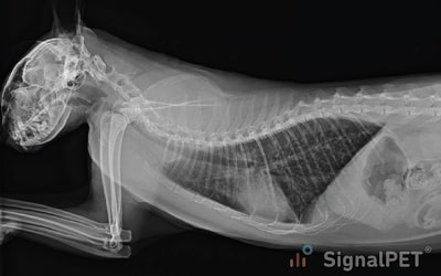
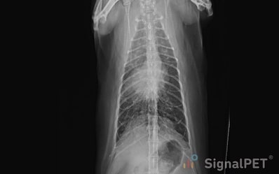
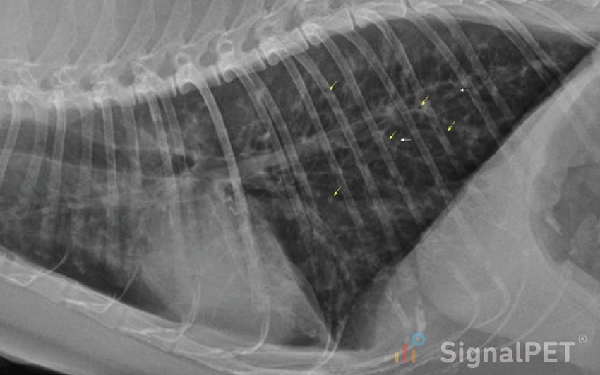
Kiwi is a 5 year old cat with moderate bronchial pattern with consolidation of the right middle lung lobe. Notice the numerous ring shadows/donuts, flattening of the diaphragm and hyperinflation of the lung lobes (line of pleural reflection extends beyond T12). Collapse of the right middle lobe will be more conspicuous in the left lateral view than in the right lateral view (effect of positional atelectasis).

Lobar collapse following chronic bronchial obstruction is observed in the right middle lobe. The collapsed right middle lobe will appear as a homogenous opacity, often triangular. The consolidated lobe can be quite small and contracted against the hilus.
Note the numerous ring shadows/donuts (yellow arrows) and tram lines (white arrows).
