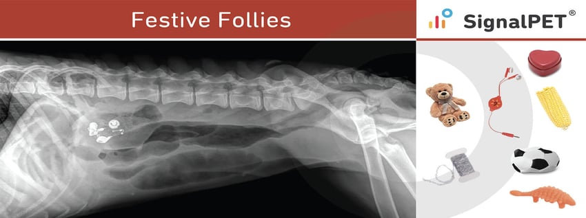
As we get to crack open the wine and tuck into the delicacies, we wanted to reflect on some common festive fumbles from this year that led the pets to emergency vet visits. This season is linked to the most common medical emergency for dogs and cats. That means anything within a paw’s reach might cause trouble, especially for curious, younger dogs. Unfortunately, this curiosity can cause significant problems such as gastric or intestinal obstruction.
Esophageal Foreign Bodies
Esophageal foreign bodies are more common in dogs than in cats and most often located at the thoracic inlet, base of the heart or just cranial to the diagram.
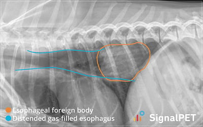
Example 1
Example of an esophageal foreign body and distended gas-filled esophagus. The foreign body is likely stuck at the level of the esophageal hiatus. Notice the tracheal stripe sign.
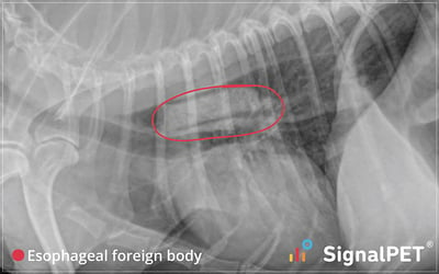
Example 2
Right lateral view of a dog with a poorly defined esophageal foreign body at the level of the thoracic inlet.
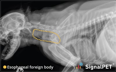
Example 3
A left lateral thoracic view of a Maltese with a esophageal foreign body at the level of the base of the heart. Notice the distended gas distended esophagus and the tracheal stripe sign.
Linear Foreign Bodies and Plication
Linear foreign bodies are string or string-like materials (tinsel, cellotape, string/plastic wrapped around meat) that get ingested by pets. They are a vet’s worst nightmare, typically necessitating immediate surgery.
Dogs and cats commonly ingest linear foreign materials, accounting for 35% of foreign bodies. It usually causes both abnormal shape and contour of the loops and an abnormal gas pattern. An “anchor” typically becomes fixed at an orad location (pylorus in dogs and under the tongue in cats), while the rest passes into the small intestine. The peristaltic action of the bowel causes the intestines to “climb” up the linear material causing the typical bunching and pleating of the affected loops. Gas becomes trapped in pockets formed by the pleats, the result being a round, sometimes crescent/comma shaped gas shapes caught in the pleats.
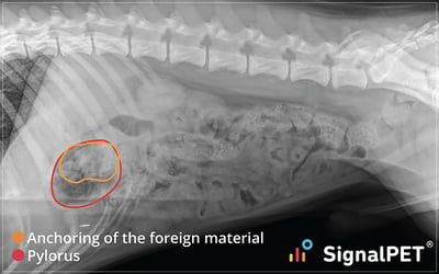
The left lateral views often support suspicion of a plication through the identification of the foreign material anchored in the pylorus and surrounded by gas.

Example 1
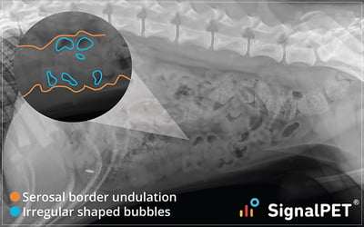
Lateral view of a Labrador Retriever with an obvious linear foreign body. The intestines are bunched in the ventral abdomen, with gas being trapped in the plications creating small irregular shaped bubbles which are normally not typical in normal bowel. Notice the relative clumping of the intestine and the pronounced serosal border undulation.
Example 2
Left lateral radiograph of a dog with a linear foreign body. The anchoring of the foreign material in the pylorus is obvious in the left lateral view due to gas being present in the pylorus.
Example 3
Lateral image of a cat with extensive intestinal linear foregin body (plication). Notice the bunching of small intestine with marked serosal undulation.
The most common foreign bodies are small toys (Dog and baby toys, balls), food (Corn cobs, pine nuts), clothing Items (Socks, underwear), bones, hair ties, rocks, dental floss, nails, fabric, rugs, etc.
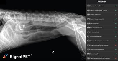
How to diagnose foreign bodies?
If you suspect that your patient might have ingested a foreign body, consider taking an abdominal X-ray, which will most likely show you metals and some other possible dense materials. Keep in mind that some other materials may not be visible. If a foreign body is not obvious on X-rays, look for characteristic intestinal gas patterns or other obstructive indicators. Looking at the GI tract by taking several timed X-rays might also be useful.

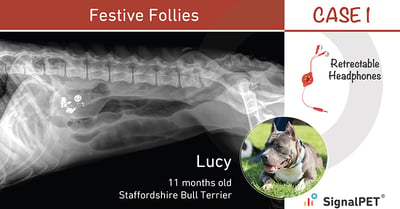
Lucy - Retractable Headphones
Kiwi is a 5 year old cat with moderate bronchial pattern with consolidation of the right middle lung lobe. Notice the numerous ring shadows/donuts, flattening of the diaphragm and hyperinflation of the lung lobes (line of pleural reflection extends beyond T12). Collapse of the right middle lobe will be more conspicuous in the left lateral view than in the right lateral view (effect of positional atelectasis).
Ronda - Heart-shaped Box
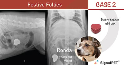
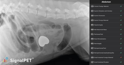
Ronda appears to have eaten a heart-shaped box. The box is visible on all views.
Suzy - Christmas Tinsel
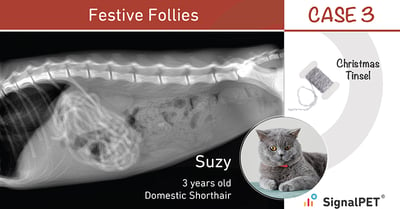
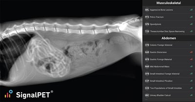
A left lateral view of Suzy, a Domestic Shorthair, that has eaten a small Christmas tinsel. Although dogs are more likely than cats to ingest foreign bodies, certain items, such as string (and Christmas Tinsel), can be irresistible to some cats.
Murphy - Toy teddy bear
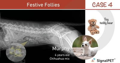
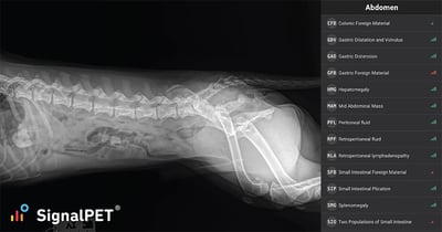
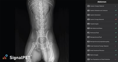
Ventrodorsal view of a Chihuahua mix with an intestinal foreign body. Close examination reveals the radiopaque structure of what appears to be a teddy bear.
Bubble - Toy Dinosaur
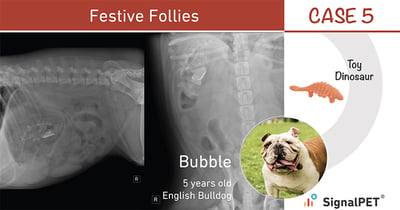
A right lateral and ventrodorsal view in an English BullDog which appeared to have ingested a toy dinosaur. Notice the gastric distension in the right lateral view and the distended population of intestinal loops (more evident in the ventrodorsal view).
Baileys - Deflated Toy Ball
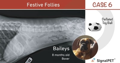
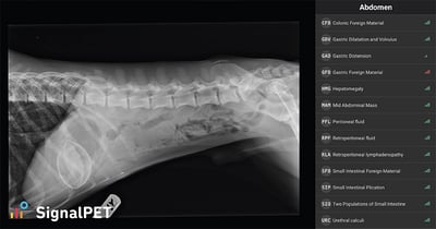
Right lateral view of a Boxer which appears to have swallowed a deflated plastic ball. Notice the radiopaque border and radiolucent gas center.
Max - Corn Cob
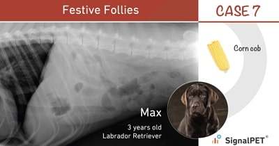
Left lateral and Ventral dorsal view of a dog that swallowed a corn-cob. Note the typical gas patterns noted as well as the distended population of intestinal loops.