Your patient is ready and waiting for you, anesthetized on the table, your technicians are waiting for direction, and you have an anxious pet parent at home waiting for updates. How do you ensure your treatment plan for this animal is correct and the patient will get the care it deserves? The answer is dental radiography.
Dental radiography is fast becoming the standard of care as part of a COHAT (comprehensive oral health assessment and treatment). The AAHA Guidelines Task Force strongly recommends intraoral radiographs for all dental patients. Studies have found that an oral exam alone does not reveal the full extent of dental disease. In dogs, up to 28% of teeth that appear normal on oral exams may be revealed to have pathology on radiographs. In cats, this percentage was much higher at 42%. With more than half of the tooth lying under the gumline, these statistics make sense.
The question naturally follows…
How do you get the most out of practicing dental radiography?
First, you must obtain quality images of the full mouth. In a cat or small dog, this would require 10-12 images. In a larger dog, 18 images may be required. Maxillary teeth will generally need 4 images on each side plus an additional occlusal image of the incisors. Mandibular teeth will generally need 3 images on each side plus an additional occlusal image of the incisors. Each tooth should only require 1 image, with individual images containing 4-8 teeth. Angle, patient positioning, and exposure all factor into the quality of these images.
Next comes interpretation of the radiographs, and SignalSMILE is available to assist. After acquisition of dental radiographs, the images are submitted to SignalSMILE. While your technicians are scaling and polishing, probing and charting, SignalSMILE is working on your images individually, uploading and analyzing, processing and reporting. Since AI reads image by image, you are able to receive results on individual images in as little as 3 minutes.
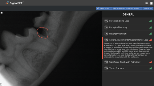
Then the review of the radiographs becomes the next important step. Test results from SignalSMILE in conjunction with your dental charting and oral exam will support you in developing a comprehensive treatment plan. SignalSMILE’s range of dental tests recognize the most common pathologies in our veterinary patients. Within the report provided on the website, you will see these pathologies show up as five separate tests: Periapical Lucency, Severe Attachment Loss, Furcation Loss, Resorptive Lesion, and Tooth Fracture. Green bars indicate a normal result, red bars indicate an abnormal result. Within each image with a pathology or pathologies identified, an outline will indicate the area of concern.
Not only can the results assist in determining your treatment plan, the SignalSMILE report can be viewed as a PDF and shared with the pet parent. This record can serve as a supportive document for the treatments you are recommending. Seeing the work completed and tests in a concise report, your client will better understand and recognize the value of work done, and will be more likely to comply with recommendations for treatment.
Lastly, if the treatment requires performing extractions, endodontic procedures, or other surgical procedures, your utilization of additional dental radiography is necessary. The American Veterinary Dental College and AAHA guidelines recommend post-operative dental radiographs. SignalSMILE is also available to assist with these images. Submit images taken as part of your original study, and SignalSMILE will report on these as well.
To learn more about SignalSMILE, or get a demo, click here.