Patient History
Presumed trauma (hit by car) and left pelvic limb lameness
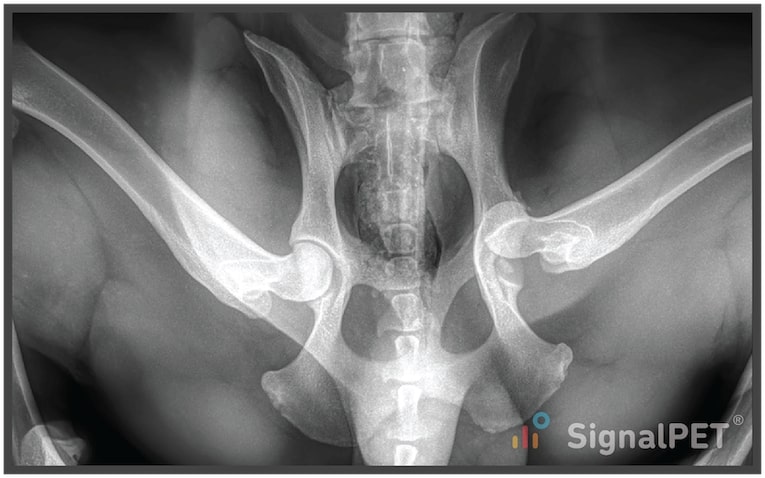
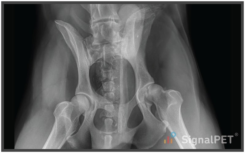
Radiographs
What’s Abnormal about this Case?
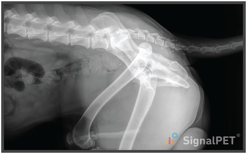
There is an obvious lesion here. On the lateral image, you can see that one hip is luxated craniodorsally. A quick glance at the orthogonal projection confirms it is the left hip.
Now what?
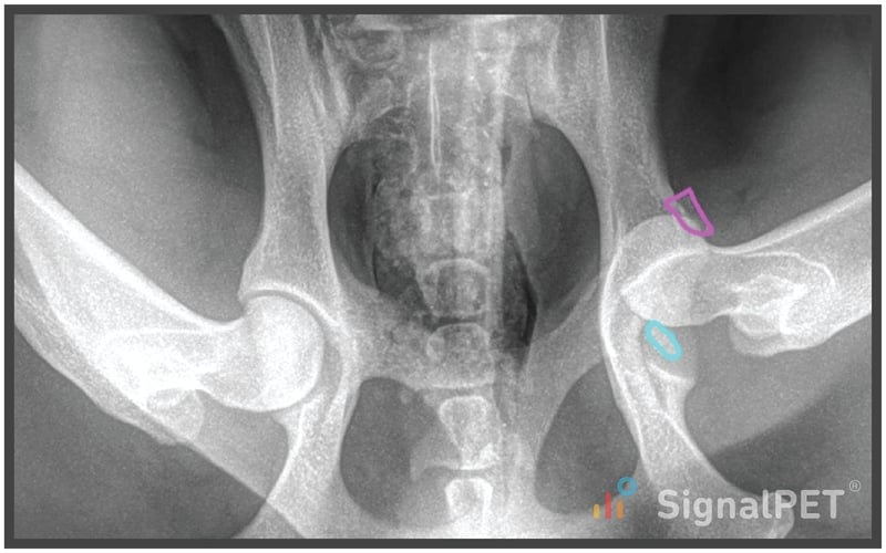
Satisfaction of search is a concept that says when we are looking for something in a radiograph, we stop looking for anything else once we find it. So in this case, the dog is limping on the left pelvic limb, so we might be inclined to stop scrutinizing the radiograph. Don’t do it! Keep looking.
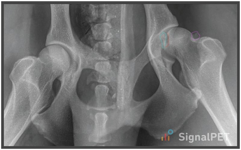
When we zoom in on the VD radiograph, we see two small mineral bodies around the left hip. One (blue) appears to be within the hip joint, while the other (pink) is adjacent to the acetabular rim. Where are they coming from? Are they important?
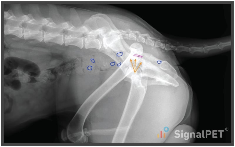
When we look on the lateral projection to try to get a better idea of where these bodies are, there is a common anatomic structure that foils our plans.
The colon!
The orange arrows indicate the empty acetabulum. Maybe there is a fragment dorsally (pink), but it looks like all the other mineral fragments (blue) in the feces. In addition to characterizing the mineral bodies, we also want to assess for long bone fractures, sacroiliac luxations, vertebral fracture/luxations, and abdominal trauma like bladder rupture and hemoabdomen (peritoneal effusion).
Radiographs post-reduction answer the question about where one of the fragments came from.
When we zoom in, there is a concave defect in the head of the femur (red) and the mineral body is next to it (blue). During hip luxation, all supporting soft tissues are torn including muscles, joint capsule, and the ligament of the head of the femur.
In this case, at the level of the normal fovea capitus, there is an avulsion fracture. This matters because the intra articular mineral body will likely serve as a cause of chronic lameness and osteoarthritis if not removed.
Other causes of mineral fragments during hip luxation include acetabular chip fractures and femoral neck avulsion fragments from the joint capsule attachment. The other fragment (pink) is likely from the femoral neck, given its location post reduction.

Plans / Tips
Remember to systematically evaluate all of the radiographs in a study, don’t just stop when you find your answer. Sometimes, small findings have big clinical implications, like this canine appendicular bone fracture case!