Patient History
Unknown.
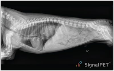
Radiographs
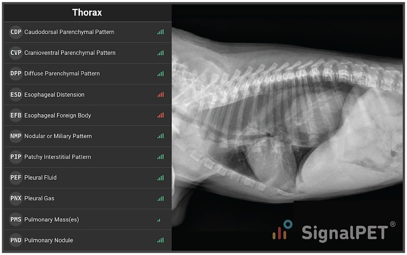
Radiology Test Results

What’s Abnormal about this Case?
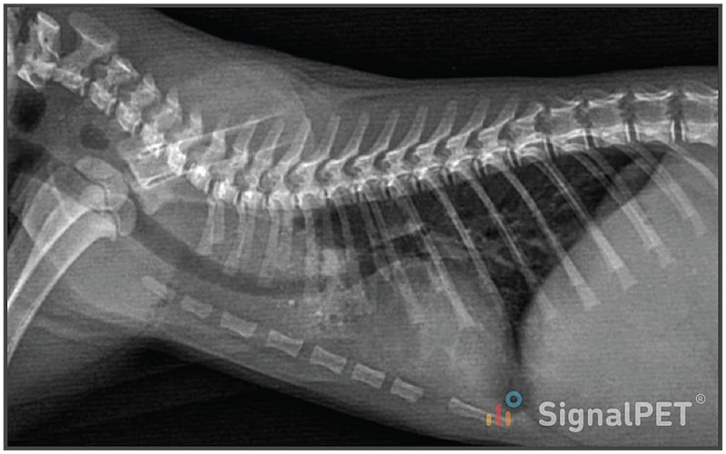
In this week’s case, the lesion is not subtle, but the disease is uncommon enough to warrant discussion. SignalRAY® identified Esophageal Distension and Esophageal Foreign Body with full confidence.
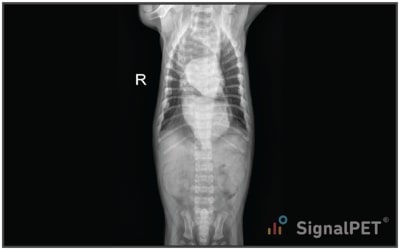
Next, we confirm the findings on the VD projection. In this case, the first thing that catches one's attention is the “two heart” effect. The cardiac silhouette is outlined in orange, while the blue outlines the stomach displaced into the distended esophagus. Again, the red arrow indicates the region where the stomach ought to be.
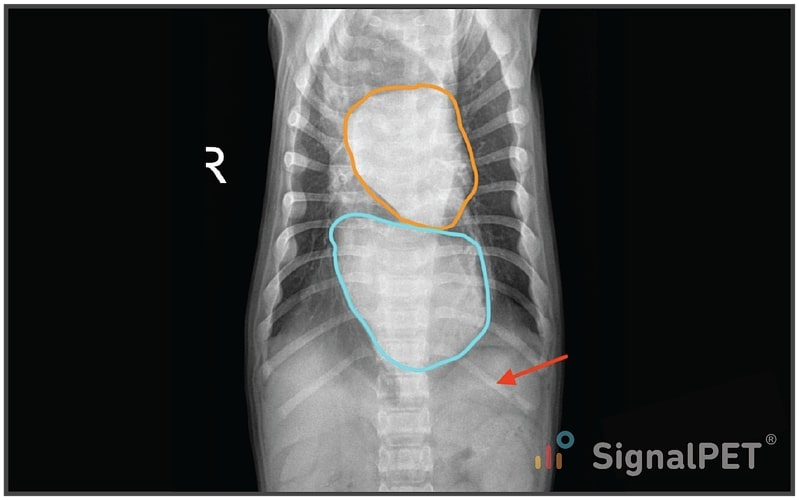
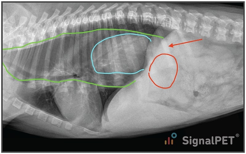
These findings are consistent with a Gastroesophageal Intussusception. This is a rare condition of unknown etiology, although an association with esophageal dysmotility is suggested. Most commonly, the condition occurs in puppies post weaning until a few months of age; German Shepherds are over represented. Typically, the patients present with acute onset, severe regurgitation and vomiting. Emergency surgical intervention is typically advocated for, although the condition can be intermittent.
Plans / Tips
When presented with a pediatric patient with regurgitation post weaning, rightly, your mind should jump to vascular ring anomaly (as pictured below), congenital megaesophagus, esophageal foreign body, and dysphagia. This case illustrates another important differential diagnosis to keep in mind and the importance of radiographs in the diagnosis. Unlike vascular ring anomalies and congenital megaesophagus, emergency surgery is indicated for gastroesophageal intussusception, which is why radiographs are indicated upon presentation.
Here is a link to an open source journal article about treating the condition endoscopically.