Patient History
Unknown - presumed vomiting
Radiographs
.jpg?width=400&name=cotw6-10%20(1).jpg)
Radiology Test Results
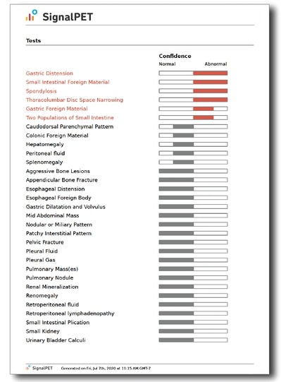

What’s Abnormal about this Case?
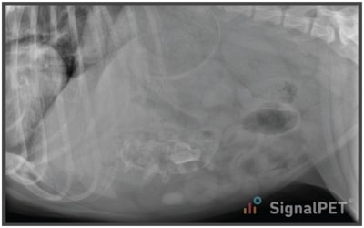
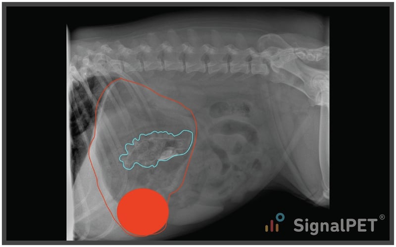
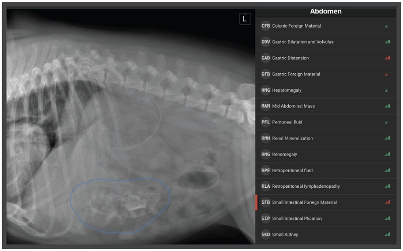
Let’s start with the right lateral projection (RLAT).
The first thing we note is the irregularly shaped, soft tissue-mineral opaque foreign body (cyan). Now the question is, where does that thing live.
The stomach is moderately distended (red) and since this is an RLAT, the pylorus contains fluid (red circle). Looks like the foreign body is floating around in the stomach with some other granular ingesta and fluid. Some of the intestinal segments are a bit dilated, but I am not sure which are colon yet. Serosal detail looks good. He’s a bulldog, so his vertebrae are malformed, but nothing else is super exciting.
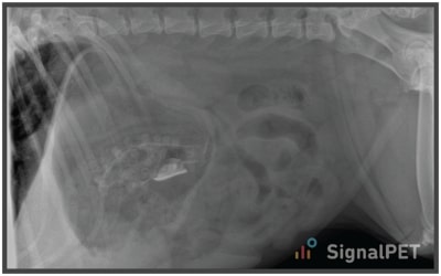
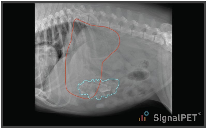
Next, I like to hop to the left lateral (LLAT) since that is my preferred projection for gastrointestinal foreign bodies. That’s because gas should move into the pylorus (if the stomach has any gas in it) and outline any foreign body wedged in the pylorus.
Here we see the foreign body again, but it appears to be projecting caudal to the stomach. Now, I switch my thinking to an intestinal foreign body and maybe even one in the pyolorduodenal junction.
In this case, it’s not outlined by gas because the stomach only contains frothy material and soft tissue opacity.
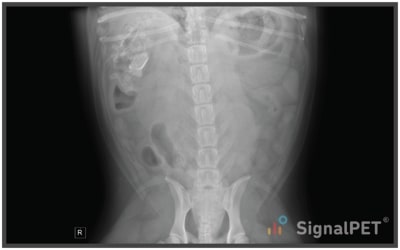
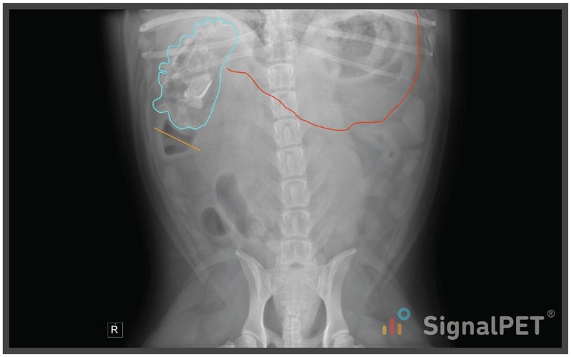
When we evaluate the VD projection, we see the foreign body again at the level of the pyloroduodenal junction with segmental enlargement of the duodenum (orange).
Now, in hindsight, if we look at the RLAT again, we can see the foreign body within the duodenum (orange).
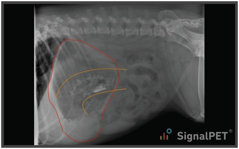
The reason the location of the foreign body matters is based on treatment options. If it’s in the stomach you could induce emesis or scope it out, but if it’s scooched out into the duodenum, it’s off to surgery.
If you are looking for a gastrointestinal obstruction, what radiographs should you take?
There is a recent paper (Mavromatis, et al. Vet Radiol Ultrasound 2018 Jul;59(4):381-386. doi: 10.1111/vru.12611) that found 2 views as good as 3 views for identifying obstructions, and in that case they used RLAT and VD vs RLAT, LLAT, and VD. However, I still prefer to have a LLAT because of visualization of the pyloroduodenal junction.
This case illustrates nicely some of the SignalRAY® results. If you open the app, you’ll see both gastric and small intestinal foreign material have red bars. If you click on Gastric Foreign Material you’ll see the RLAT thumbnail is red, indicating the abnormal sign is on that image. If you then click on Small Intestinal Foreign Material you’ll see the LLAT and VD thumbnails are red. Remember, AI looks for signs on individual images, so it doesn’t have the information from the LLAT and VD to apply back to the RLAT.
.jpg?width=800&name=cotw6-10%20(1).jpg)
Plans / Tips
My 2 cents when it comes to vomiting cats and dogs - take at least an LLAT and VD.
If that answers the question, great, but sometimes adding the RLAT offers one more view that adds confidence to your decision.